Join the Machine Learning in Medical Imaging Consortium (MaLMIC) for an opportunity to network on machine learning
Monthly Virtual Forum Series on Zoom
Friday, November 19, 2021
3:30 to 5:30 p.m. EST
Dr. Emily Conant, a radiologist from the University of Pennsylvania, gave the lead-off talk and presented the clinical problems and opportunities where machine learning could make significant impact. She was followed by Professor Nico Karssemeijer from Radboud University in the Netherlands. He has developed important algorithms for breast density and is implementing AI approaches to detection of breast cancer. Grey Kuling, PhD candidate from Sunnybrook Research Institute gave the third talk covering the more technical approach of their research work. The talks were followed by discussion focused on lessons learned, opportunities to collaborate and sharing of resources.


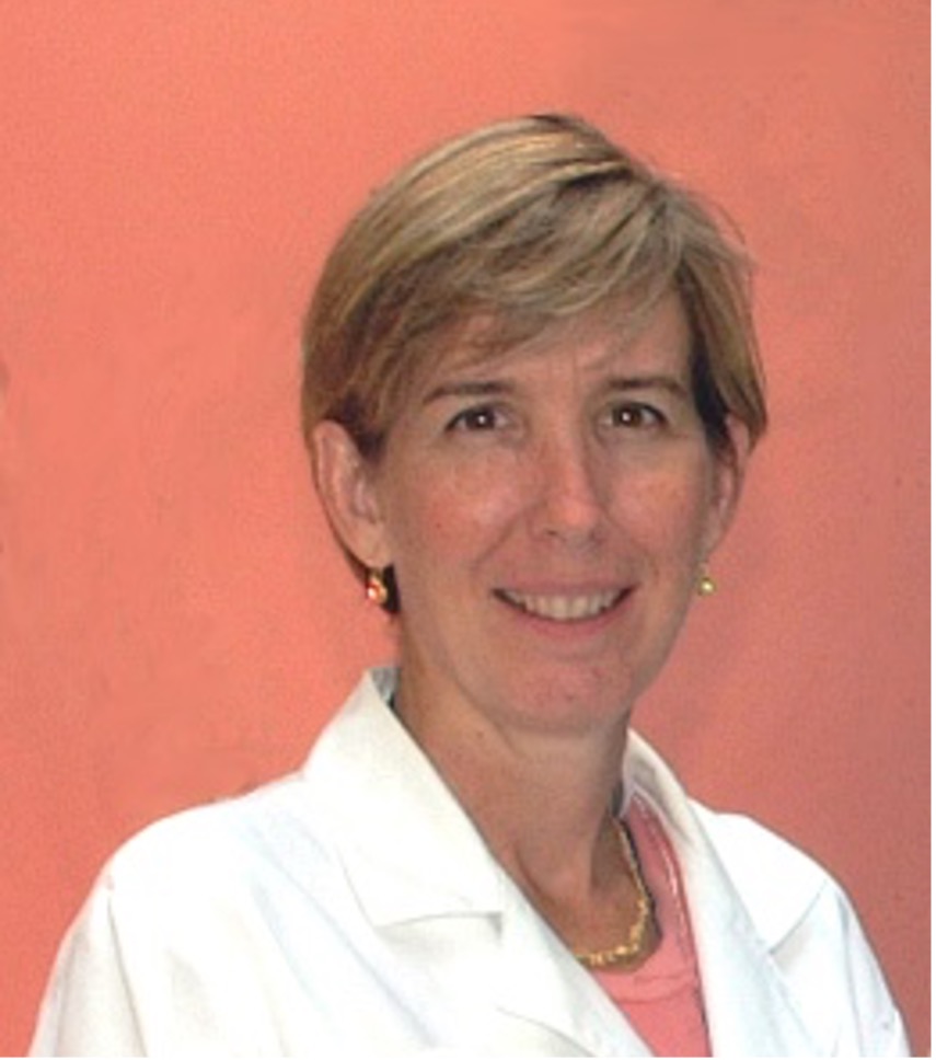
Artificial Intelligence Applications in Breast Cancer Screening: A Clinical Perspective
Emily Conant, MD
Professor of Radiology, University of Pennsylvania Perelman School of Medicine
Talk summary: Emily discussed the various challenges that we face in mammographic screening that incorporating AI applications may help with. These areas included:
- Interpretative Tasks
- improvements in patient outcomes by decreasing the variability of reads as well as improving both sensitivity and specificity of reads
- Non-interpretative Tasks
- improvements in efficiency of reads (i.e., using AI to aid a single radiologist read instead of double reading by 2 radiologists) and the use of AI to “triage” cases by complexity
- improvements in “Precision Screening” by incorporating image data in breast cancer risk assessment algorithms


Application of artificial intelligence for early detection of breast cancer
Nico Karssemeijer, PhD
Professor, Radboud University, Nijmegen, NL
Talk summary: Nico discussed methods to compare standalone performance of AI for reading screening mammograms with radiologists and issues that complicate such comparisons. The state of art was illustrated with examples. Use of AI as an autonomous reader can lead to more efficient and effective workflows in breast screening screening practice, reducing the need for a demanding double reading process. AI may also support implementation of personalised breast screening, where women with an elevated risk are offered supplemental screening with more sensitive modalities such as (abbreviated) MRI. Tools to support radiologists with the interpretation of breast MRI are being developed as well.


